
Введение
Пролапс – выпадение слезной железы третьего века – является распространенной патологией, встречающейся преимущественно у молодых собак брахицефальных, крупных и гигантских пород. Однако данное заболевание может наблюдаться и у собак иных пород и возрастов, а также у кошек (фото 1).Этиология пролапса у собак остается не до конца выясненной, но предполагается, что основными факторами являются породная предрасположенность (генетические факторы), анатомические особенности (мелкая орбита у брахицефалов) и рыхлость соединительной ткани третьего века1.
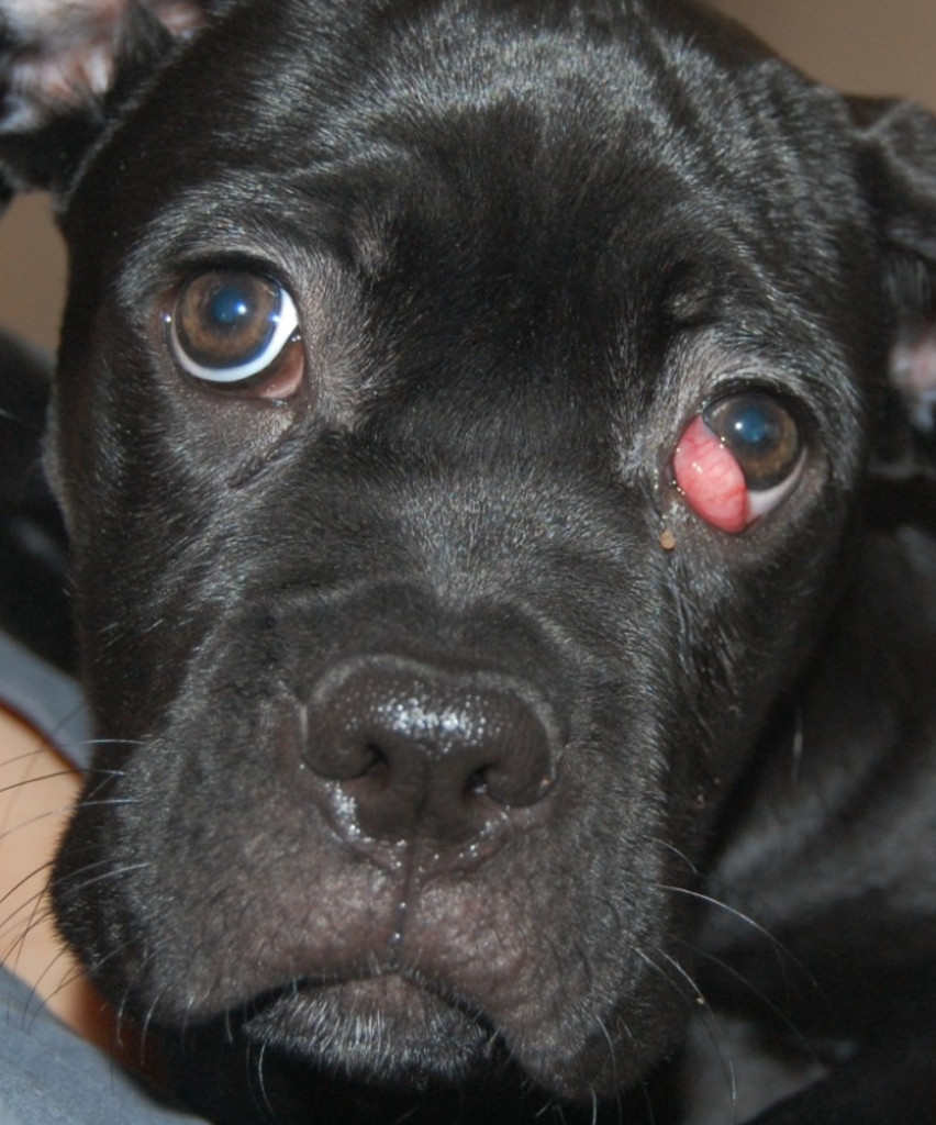
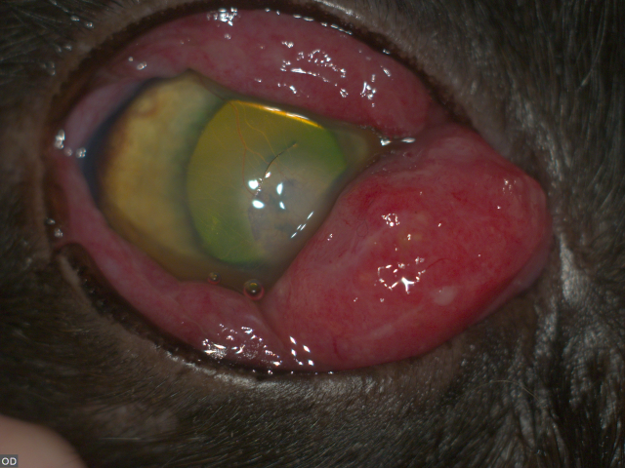
Удаление слезной железы при ее пролапсе не проводится, поскольку слезная железа третьего века выделяет до 30% всей слезной жидкости, омывающей глаз, и ее удаление может стать фактором, провоцирующим или усугубляющим течение сухого кератоконъюнктивита1,9. Единственным показанием к удалению слезной железы является наличие новообразования (чаще всего аденокарциномы, реже – плоскоклеточной карциномы), которое необходимо дифференцировать от пролапса (фото 2). Критериями могут служить следующие факторы:
- Возраст животного: у животных старшей возрастной группы (особенно у кошек) новообразования встречаются чаще, чем у молодых особей.
- Анамнез: постепенный рост тканей характерен для новообразований, в то время как пролапс обычно возникает внезапно.
- Внешний вид и текстура тканей: при новообразованиях ткани имеют бугристую, неровную поверхность и плотную консистенцию, а при пролапсе они гладкие, упругие и однородные.
- Результаты гистологического исследования биоптата: при подозрении на новообразование рекомендуется провести инцизионную биопсию и направить полученный материал на гистологическое исследование.
Существует множество методов для коррекции выпадения слезной железы, включая ручное вправление под местной анестезией, хирургические техники с фиксацией железы или формированием кармана, а также различные комбинации и модификации этих техник.
Техники вправления выпавшей слезной железы третьего века
Ручное вправление может применяться для временной коррекции пролапса слезной железы при свежем первичном выпадении. Эта процедура наиболее эффективна на ранних стадиях, до развития отека и воспаления железы.Техника выполнения: после применения местной анестезии (двукратная или трехкратная инстилляция препарата «Инокаин» в конъюнктивальный мешок) выполняется смещение третьего века из медиального угла глаза путем мягкого надавливания на глазное яблоко через верхнее веко в дорсолатеральной части (это позволит визуализировать свободный край третьего века, расположенный под выпавшей железой). Свободный край третьего века захватывают атравматичным пинцетом по Грефе и тянут в дорсолатеральном направлении, выворачивая веко в анатомически правильное положение. Параллельно с этим можно надавить на поверхность выпавшей слезной железы ватной палочкой, смещая ее в конъюнктивальный свод вентромедиально.
Эффект ручного вправления может быть кратковременным (от нескольких часов до нескольких дней), а иногда длительным (на протяжении нескольких месяцев).
Хирургические техники вправления
Метод кармана по Моргану, впервые описанный Морганом с соавторами (Morgan R. V., et al.) в 1993 году5, до сих пор широко применяется в клинической практике.Техника выполнения: после введения общей анестезии пациента располагают в боковом положении, верхнее и нижнее веки удерживают векорасширителем, третье веко фиксируют за складку конъюнктивы с пальпебральной поверхности двумя-тремя маленькими гемостатическими зажимами типа «москит», чтобы расправить третье веко и полноценно визуализировать поверхность слезной железы. Затем выполняются два линейных разреза конъюнктивы вдоль переднего края слезной железы (в месте перехода ее поверхности в плоскую часть конъюнктивы, покрывающую свободную часть третьего века) и параллельно первому – вдоль задней границы слезной железы. Края разрезов соединяют между собой швом над железой, погружая ее в своеобразный карман: первый узел шовного материала (обычно ПГА 6-0) располагают на пальпебральной поверхности третьего века, после чего прокалывают веко насквозь, выводя нить на бульбарную поверхность и используют погружной шов типа «елочка». После завершения шва веко прокалывается еще раз, чтобы второй узел также был завязан на пальпебральной поверхности, предотвращая контакт шовного материала с роговицей и снижая риск образования язв и эрозий (видео 1).
Преимуществами данной методики являются простота, быстрота выполнения и низкая травматичность. К недостаткам можно отнести риск раннего рецидива из-за разрушения шовного материала до формирования рубца конъюнктивы, а также вероятность развития кисты слезной железы.
Видео 1. Метод кармана по Моргану.
Смотреть в VK
Технику «кисетного шва» или метод фиксации слезной железы к хрящу века описали Пламмер с соавторами (Plummer C. E., et al.) в 2008 году7. Суть метода заключается в использовании нерассасывающейся монофиламентной нити размером 4-0 или 3-0 для фиксации слезной железы к ножке хряща третьего века. Игла вводится в центр вертикальной части Т-образного хряща третьего века с пальпебральной поверхности на бульбарную. Затем несколькими последовательными стежками обходят по периметру железы так, чтобы шовный материал не образовал петель над конъюнктивой и не травмировал роговицу, для этого каждый последующий вкол иглы выполняют в том же месте, откуда ранее игла выходила. После обхода железы по периметру с помощью таких стежков хрящ третьего века вновь прокалывают насквозь недалеко от места первого его прокола, что позволяет вывести оба конца нити на пальпебральную поверхность третьего века, где они завязываются в узел. Натяжение нити приводит к формированию складки конъюнктивы бульбарной поверхности третьего века над слезной железой, создавая подобие «кисета», который удерживает железу на месте и предотвращает ее выпадение (видео 2).
Видео 2. Техника «кисетного шва».
Смотреть в VK
К плюсам данной техники относят простоту, быстроту выполнения, низкую травматичность, к минусам – риск рецидива из-за смещения или разрыва шовного материала, риск травмы роговицы при смещении узла.
Методика фиксации железы к надкостнице скуловой кости, которую впервые описали Касван и Мартин (Kaswan R., Martin C.) в 1985 году, а затем дополнили Касван и Стенли (Kaswan L. R., Stanley R. G.) в 1994 году11, заключается в проведении нерассасывающейся монофиламентной нити вокруг железы и через надкостницу скуловой кости вентрального края орбиты.
Для проведения процедуры выполняются два разреза: кожи нижнего века над вентромедиальным краем орбиты и конъюнктивы в центре вентрального конъюнктивального свода. Режущую иглу от монофиламентного нерассасывающегося шовного материала размером 2-0, 3-0 или 4-0 (в зависимости от размера пациента) вводят в кожный разрез, захватывая участок надкостницы на краю орбиты, и выводят из разреза вентрального конъюнктивального свода. Вторую иглу, расположенную на другом конце нити, аналогичным образом проводят через надкостницу, таким образом оба конца нити оказываются выходящими из разреза вентрального конъюнктивального свода. Далее одна из игл возвращается в конъюнктивальный разрез и вводится в толщу третьего века дорсально вдоль короткой стороны слезной железы. Выход иглы происходит через бульбарную поверхность конъюнктивы третьего века. Следующий вкол иглы осуществляется в том же месте, где был выкол, что предотвращает травматизацию роговицы петлями шовного материала; шов проводят в толще третьего века вдоль длинной стороны железы. Третий стежок выполняется в вентральном направлении вдоль второй короткой границы слезной железы, после чего игла выводится на пальпебральную поверхность третьего века в разрез вентрального конъюнктивального свода, здесь концы шовного материала фиксируются между собой узлом. Разрезы кожи и вентрального конъюнктивального свода ушиваются по мере необходимости.
Плюсы данной методики: малая травматичность для тканей железы, значительное смещение третьего века вентромедиально (может быть полезно для некоторых собак с косметической целью), минусы – более длительная операция, надежность фиксации зависит от сохранности шовного материала, ограничение подвижности третьего века.
Фиксация железы к вентральной прямой мышце, которую предложили Сапиенца с соавторами (Sapienza J. S., et al.) в 2014 году10, считается более надежной для гигантских и рыхлых пород собак.
Техника выполнения: третье веко фиксируют за складку конъюнктивы с пальпебральной поверхности двумя-тремя маленькими гемостатическими зажимами типа «москит» для обеспечения полноценной визуализации поверхности слезной железы. Скальпелем № 15 выполняют линейный разрез бульбарной конъюнктивы третьего века над слезной железой в направлении от глазного яблока к свободному краю третьего века, расширяют разрез конъюнктивы ножницами, чтобы обнажить железу. Слезную железу отделяют от хряща третьего века с помощью ножниц, а также иссекают небольшой участок основания хряща. Под точкой прикрепления вентральной прямой мышцы к глазному яблоку вводят мышечный крючок, через который проводят под мышцей нерассасывающийся монофиламентный шовный материал (например, Этилон 5-0). Иглой от этого шовного материала делают прокол вдоль слезной железы (в области наибольшей проминенции железы, параллельно свободному краю века), с помощью ватной палочки смещают слезную железу вентрально к глазному яблоку, затем, удалив ватную палочку, затягивают узел шовного материала, фиксирующий железу к прямой мышце, конъюнктиву не ушивают.
Преимуществами данной методики являются хороший косметический результат у собак гигантских пород и высокая надежность. К недостаткам можно отнести длительность операции, травматичность по отношению к тканям железы и риск рецидива при преждевременном нарушении целостности шва.
Существует множество вариаций и комбинаций методов хирургического лечения. Например, метод кармана может быть дополнен фиксацией слезной железы к надкостнице скуловой кости6, укорочением длины третьего века4 или модификацией метода кармана с фиксацией слезной железы к склере2. Также известны и другие модификации8.
Стандартный послеоперационный протокол лечения продолжительностью около 7–14 дней включает использование местного антибиотика широкого спектра действия и защитного воротника. В некоторых случаях может быть назначен местный стероид.
Осложнения, их профилактика и коррекция
Осложнения после хирургического вмешательства могут включать рецидивы (ранние и поздние), кисты слезной железы, травмы роговицы шовным материалом и воспаление.Ранний рецидив пролапса (в период до 7 дней после операции) чаще всего обусловлен самотравмированием и преждевременным разрушением или смещением шовного материала (до формирования рубца конъюнктивы), поэтому для профилактики подобных осложнений рекомендуется использование защитного воротника в течение первых 5–7 дней после операции.
Более поздние рецидивы (спустя 7 и более дней после операции) в условиях уже сформированного рубца тканей вокруг железы чаще всего связаны с нарушением техники операции, развитием сочетанных патологий хряща третьего века, значительными изменениями размеров животного и усугублением выворота нижнего века, особенно у гигантских и рыхлых пород собак, у которых операция проводится в молодом возрасте.
Любые случаи рецидива требуют вмешательства для ревизии конъюнктивального мешка, поиска причин рецидива и повторного хирургического вправления железы в нормальное анатомическое положение. Рецидив не является показанием к удалению слезной железы третьего века.
Киста слезной железы третьего века может возникать как осложнение хирургических техник вправления, при которых происходит травматизация выводных протоков железы, а также образование рубцовой ткани над железой, что препятствует нормальному дренажу слезы в конъюнктивальный мешок (например, метод кармана). Для профилактики формирования кист при использовании техники кармана по Моргану рекомендуется неполное сопоставление краев разрезов (при этом оставляют отверстия размером около 2–3 мм в области начала и конца шва бульбарной поверхности конъюнктивы третьего века) и сохранение интактной конъюнктивы на поверхности слезной железы третьего века. Однако эффективность этих мер не может быть гарантирована с абсолютной уверенностью.
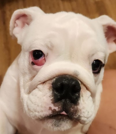
Киста обычно формируется в течение 1–3 недель после проведения операции, клинически она проявляется протрузией и утолщением третьего века. При пальпации можно идентифицировать упругую флюктуирующую область в третьем веке, при пункции которой обнаруживается жидкий прозрачный секрет (слеза). После пункции и эвакуации содержимого киста быстро заполняется в течение нескольких дней (фото 3).
Методом коррекции кист слезной железы третьего века является марсупиализация – хирургическая техника, при которой стенки кисты сшивают с конъюнктивой так, чтобы сформировать дренажное отверстие3. Процедура выполняется следующим образом: не опорожняя кисту заранее, определяют максимально проминирующую ее часть, над которой проводят разрез конъюнктивы (предпочтительно с пальпебральной поверхности третьего века) и подлежащей стенки кисты. После истечения содержимого кисты шовным материалом ПГА 6-0 или 7-0 простыми узловыми швами сшивают по кругу край стенки кисты с краем разреза конъюнктивы. Послеоперационный уход аналогичен таковому при хирургическом вправлении пролапса.
Миграция шовного материала может происходить при различных техниках вправления. Клиническими проявлениями такого состояния обычно являются блефароспазм, эпифора и эрозия или язва роговицы, чаще всего локализующаяся в проекции третьего века.
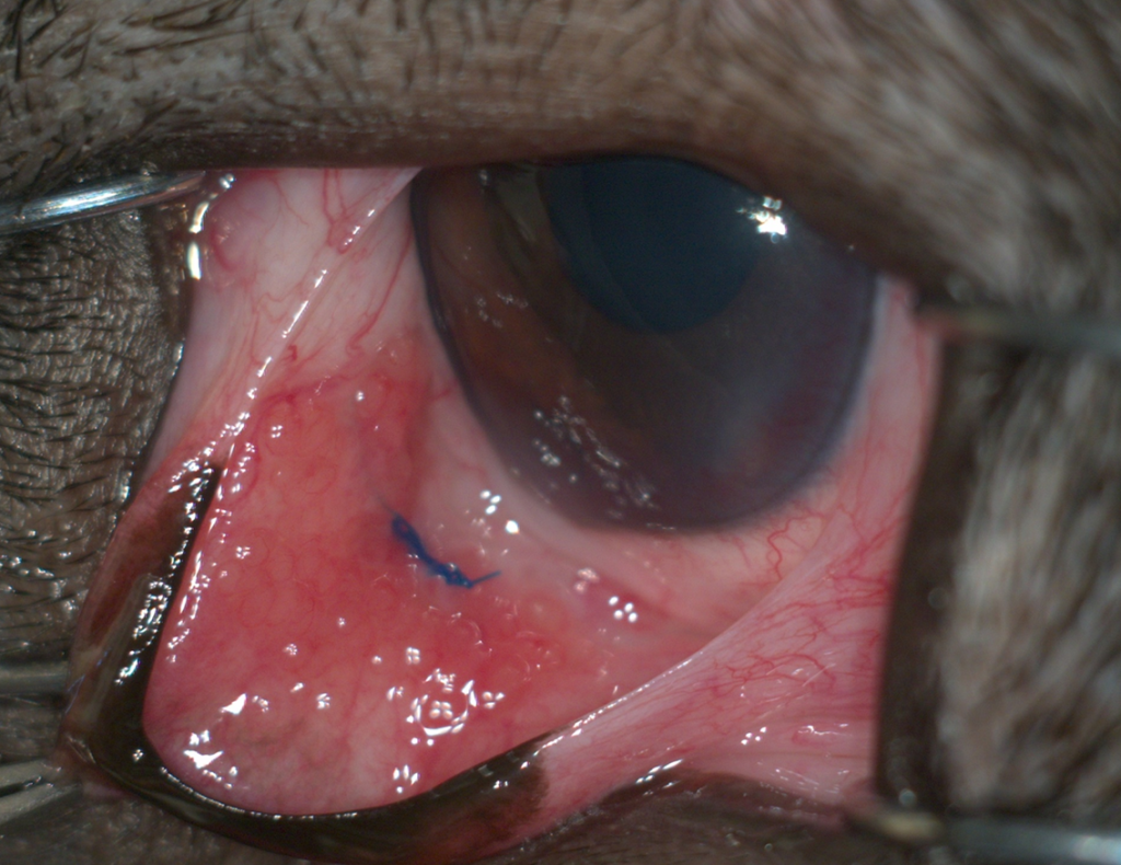
При применении техники кармана по Моргану используется рассасывающийся шовный материал, который при самотравмировании или постепенном физиологическом разрушении может выходить на поверхность конъюнктивы, в таком случае шовный материал можно удалить с применением местной анестезии (при условии спокойного поведения животного) или при ревизии конъюнктивального мешка с применением общей анестезии в иных случаях. Если к моменту необходимости удаления шовного материала уже сформировался стабильный рубец в тканях, то устранение материала не увеличит риск рецидива пролапса. Миграция нерассасывающегося шовного материала при использовании техники «кисетного шва» может привести к рецидиву пролапса, особенно при смещении шва, что не обязательно связано с травмой роговицы шовным материалом. Также возможны повреждения роговицы петлями шовного материала или его узлом, что вызывает серьезные повреждения роговицы и болевой синдром (фото 4).
Для устранения миграции нерассасывающегося шовного материала может потребоваться повторное хирургическое вмешательство по вправлению слезной железы, поскольку простое удаление шовного материала оставляет железу без фиксации.
Воспаление тканей конъюнктивы и/или слезной железы как осложнение хирургического вправления встречается редко. Оно может быть обусловлено использованием неподходящего или загрязненного шовного материала, неправильным выбором диаметра шовного материала, несоблюдением принципов асептики/антисептики, а также нарушением техники операции (грубые манипуляции с тканями, размозжение тканей), самотравмированием или индивидуальной реакцией организма животного на рассасывающийся шовный материал.
При наличии избыточного отека и протрузии третьего века, гнойных выделений из конъюнктивального мешка в послеоперационном периоде рекомендуются ревизия конъюнктивального мешка, устранение шовного материала (при его миграции, несостоятельности швов), а также терапевтические методы контроля (местные и/или системные противовоспалительные препараты и антибиотики).
Заключение
Пролапс слезной железы третьего века часто встречается в практике ветеринарного офтальмолога и требует своевременного хирургического лечения – вправления железы. Описанные в литературе техники хирургического вправления разнообразны и могут быть применены в зависимости от показаний каждого конкретного пациента и по предпочтению хирурга.Осложнения хирургических техник и даже рецидивы не должны стать показанием к удалению здоровой ткани слезной железы, а могут быть скорректированы описанными в данной статье методами.
Список литературы:
- Giuliano E., Diseases and Surgery of the Canine Lacrimal Secretory System, in Veterinary Ophthalmology, 5th ed., ed. K. Gelatt, B. Gilger, and T. Kern (Wiley and Sons, 2013) 922936–937.
- Guionnet A. and Weverberg F. Surgical Correction of Prolapse of Nictitating Membrane Gland Using a Variant of the Pocket Technique: A Retrospective Study on 101 Dogs and 126 Eyes, Veterinary Ophthalmology, 29, no. 1, 2026; e70031, ссылка.
- Lima T. B. & Martins T. B. & Junior D. C. & Silva R. A. & Sousa D. M. F. Marsupialization for the treatment of nictitating membrane cyst in a dog: case report. Arquivo Brasileiro de Medicina Veterinária e Zootecnia, 72:749–753, 2020. 10.1590/1678-4162-11033.
- Michel J., Lazard P., Vigan M., Albaric O. Treatment of prolapsed gland and cartilage deformity of the nictitating membrane with pocket technique and chondrectomy alone, or combined with a wedge conjunctivectomy: 132 dogs (1998-2018). Vet Ophthalmol, 23(2):305–313, Mar 2020. doi: 10.1111/vop.12727.
- Morgan R. V., Duddy J. M., McClurg K. Prolapse of the gland of the third eyelid in dogs: a retrospective study of 89 cases (1980 to 1990). Journal of the American Animal Hospital Association, 29:56–60, 1993.
- Multari, Domenico & Perazzi, Anna & Contiero, Barbara & Mattia, Giada & Iacopetti, Ilaria. Pocket technique or pocket technique combined with modified orbital rim anchorage for the replacement of a prolapsed gland of the third eyelid in dogs: 353 dogs. Veterinary ophthalmology, 19, 2016. 10.1111/vop.12286.
- Plummer C. E., Kallberg M. E., Gelatt K. N. et al. Intranictitans tacking for replacement of prolapsed gland of the third eyelid in dogs. Veterinary Ophthalmology, 11:228–233, 2008.
- Premont J. E., Monclin S., Farnir, F. et al. Perilimbal Pocket Technique for Surgical Repositioning of Prolapsed Nictitans Gland in Dogs. Veterinary Record, 171:247–252, 2012.
- Saito A., Watanabe Y., and Kotani T. Morphologic Changes of the Anterior Corneal Epithelium Caused by Third Eyelid Removal in Dogs, Veterinary Ophthalmology, 7:113–119, 2004.
- Sapienza J. S., Mayordomo A., Beyer A. M. Suture anchor placement technique around the insertion of the ventral rictus muscle forthe replacement of the prolapsed gland of the third eyelid in dogs: 100 dogs. Veterinary Ophthalmology, 17:81–86, 2014.
- Stanley R. G., Kaswan L. R. Modification of the orbital rim anchorage method for surgical replacement of the gland of the third eyelid in dogs. Journal of the American Veterinary Medical Association, 205:1412–1414, 1994.
 vetpeterburg
 Ветеринарный Петербург